
Medical imaging has transformed healthcare by enabling clinicians to diagnose diseases accurately, monitor treatment progress, and perform minimally invasive procedures. Among all imaging modalities, ultrasonography has emerged as one of the most versatile, cost-effective, and widely used diagnostic tools across almost every medical specialty.
From emergency departments and intensive care units to obstetrics, cardiology, nephrology, surgery, orthopedics, and primary healthcare, ultrasound plays an indispensable role in patient management. The demand for physicians skilled in ultrasonography is growing rapidly as hospitals, diagnostic centers, and clinics increasingly rely on ultrasound for early diagnosis and improved patient outcomes.
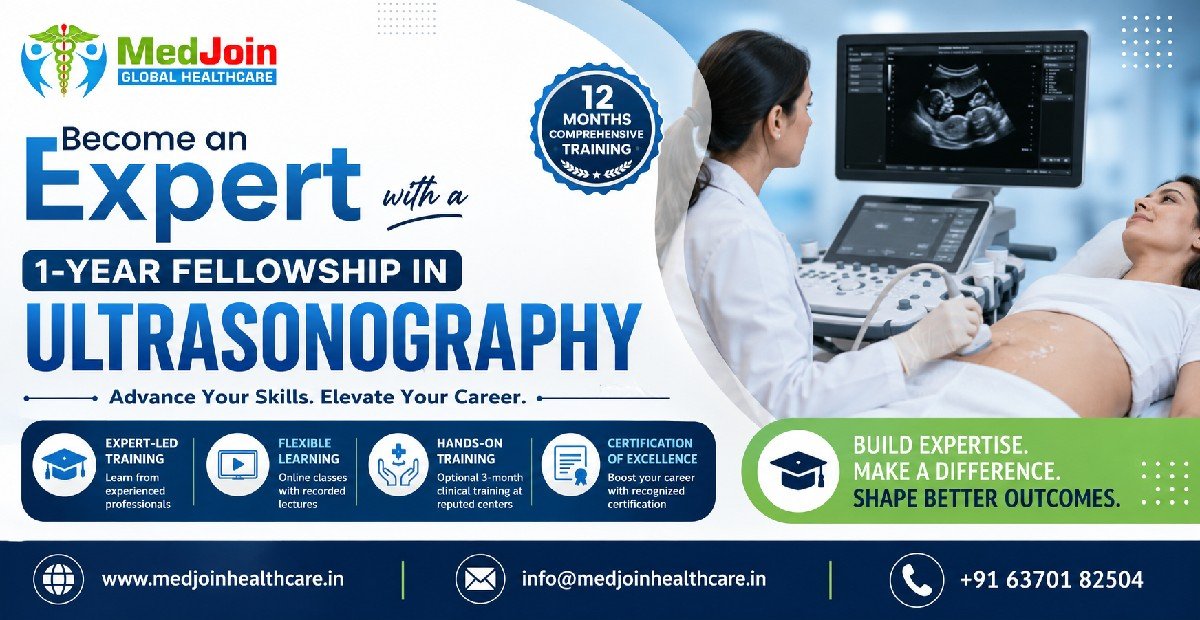
For doctors looking to strengthen their clinical expertise and stay ahead in today's competitive healthcare landscape, a 1-Year Fellowship in Ultrasonography provides a structured pathway to acquire advanced knowledge, develop practical skills, and enhance career opportunities.
Whether you are an MBBS graduate, postgraduate resident, general physician, family physician, emergency physician, or specialist, this fellowship equips you with the competence needed to integrate diagnostic ultrasound into routine clinical practice.
Why Ultrasonography Matters in Modern Healthcare
Ultrasonography has revolutionized diagnostic medicine because it offers real-time visualization of internal organs without exposing patients to ionizing radiation.
Unlike CT scans or X-rays, ultrasound is:
- Safe
- Non-invasive
- Radiation-free
- Cost-effective
- Portable
- Repeatable
- Bedside-friendly
These advantages have made ultrasound the preferred first-line imaging investigation in numerous clinical situations.
Today, clinicians use ultrasound for:
- Abdomen evaluation
- Liver diseases
- Gall bladder pathology
- Kidney disorders
- Urinary tract diseases
- Pregnancy assessment
- Gynecological disorders
- Cardiac assessment
- Vascular imaging
- Musculoskeletal injuries
- Thyroid examination
- Breast imaging
- Emergency trauma evaluation
- Critical care monitoring
- Pediatric imaging
- Neonatal imaging
- Guided procedures
The growing dependence on ultrasonography has created a significant need for physicians with advanced ultrasound expertise.
Growing Demand for Ultrasonography Specialists
Healthcare systems across India and globally are witnessing tremendous growth in diagnostic imaging services.
Several factors are driving this demand:
Increasing Burden of Chronic Diseases
Diseases such as:
- Diabetes
- Hypertension
- Fatty liver disease
- Chronic kidney disease
- Heart disease
- Cancer
require frequent imaging for diagnosis and follow-up.
Ultrasound remains the preferred initial imaging modality for many of these conditions.
Expansion of Preventive Healthcare
Preventive medicine emphasizes early disease detection.
Routine health check-ups now commonly include ultrasound examinations for:
- Liver
- Kidney
- Thyroid
- Pelvis
- Prostate
- Breast
- Carotid arteries
As preventive healthcare expands, ultrasound-trained physicians become increasingly valuable.
Growth of Point-of-Care Ultrasound (POCUS)
Modern medicine is shifting toward bedside imaging.
Instead of sending patients to radiology departments, clinicians now perform ultrasound themselves during patient examination.
This approach is known as:
Point-of-Care Ultrasound (POCUS).
POCUS is widely adopted in:
- Emergency Medicine
- Internal Medicine
- ICU
- Critical Care
- Nephrology
- Cardiology
- Anesthesiology
- Family Medicine
Doctors trained in ultrasonography can make faster and more accurate clinical decisions.
Rising Number of Diagnostic Centers
India has experienced rapid growth in:
- Imaging centers
- Multispecialty hospitals
- Corporate hospitals
- Medical colleges
- Day-care centers
- Dialysis centers
These facilities require qualified ultrasound professionals to meet increasing patient volumes.
What is a 1-Year Fellowship in Ultrasonography?
A 1-Year Fellowship in Ultrasonography is an advanced professional training program designed to provide doctors with comprehensive theoretical knowledge and practical skills in diagnostic ultrasound.
The fellowship covers:
- Ultrasound physics
- Instrumentation
- Image optimization
- Scanning protocols
- Normal anatomy
- Pathological findings
- Doppler imaging
- Color Doppler
- Vascular ultrasound
- Emergency ultrasound
- Procedure guidance
- Clinical applications
The program bridges the gap between theoretical learning and real-world clinical practice.
Participants learn systematic approaches to image acquisition, interpretation, and reporting while understanding the strengths and limitations of ultrasound in patient care.
Why Doctors Should Learn Ultrasonography
The role of physicians is evolving.
Modern clinicians are expected to combine clinical examination with bedside imaging.
Ultrasound significantly improves diagnostic accuracy.
For example:
Instead of relying solely on physical examination, physicians can immediately confirm findings such as:
- Gallstones
- Ascites
- Pleural effusion
- Deep vein thrombosis
- Hydronephrosis
- Cardiac tamponade
- Abdominal aortic aneurysm
- Pregnancy
- Liver cirrhosis
This improves patient care while reducing unnecessary investigations.
Benefits of Learning Ultrasonography
1. Better Clinical Decision Making
Real-time imaging enables physicians to diagnose diseases quickly.
This reduces delays in treatment and improves outcomes.
2. Improved Patient Safety
Ultrasound avoids radiation exposure, making it safe for:
- Pregnant women
- Children
- Neonates
- Critically ill patients
3. Higher Diagnostic Confidence
Clinicians gain confidence by correlating:
- History
- Physical examination
- Laboratory findings
- Ultrasound imaging
4. Expanded Scope of Practice
Doctors can independently perform many bedside imaging procedures without waiting for radiology appointments.
5. Better Career Opportunities
Hospitals increasingly prefer physicians with ultrasound expertise.
Additional imaging skills strengthen CVs and improve employability.
Clinical Applications of Ultrasonography
One of ultrasound's greatest strengths is its versatility.
It supports diagnosis across numerous specialties.
Internal Medicine
Applications include:
- Liver diseases
- Fatty liver
- Cirrhosis
- Splenomegaly
- Kidney diseases
- Hydronephrosis
- Bladder pathology
- Ascites
- Pleural effusion
Emergency Medicine
Emergency physicians use ultrasound for:
- FAST examination
- Trauma evaluation
- Shock assessment
- Cardiac arrest
- Pneumothorax
- Hemoperitoneum
- Aortic aneurysm
- Bedside procedures
Rapid diagnosis can be life-saving.
Obstetrics and Gynecology
Ultrasound is essential for:
- Pregnancy dating
- Fetal growth monitoring
- Placental localization
- Ectopic pregnancy
- Ovarian cysts
- Uterine fibroids
- Infertility assessment
Nephrology
Ultrasound assists in:
- Kidney size evaluation
- Chronic kidney disease
- Obstruction
- Renal cysts
- Renal masses
- Dialysis catheter guidance
- Kidney biopsy guidance
Cardiology
Cardiac ultrasound evaluates:
- Ventricular function
- Valvular disease
- Pericardial effusion
- Cardiomyopathy
- Congenital heart disease
Critical Care
ICU physicians perform bedside ultrasound to assess:
- Lung pathology
- Pleural effusion
- Cardiac function
- Fluid responsiveness
- Central venous access
- Deep vein thrombosis
Pediatrics
Applications include:
- Neonatal brain ultrasound
- Hip ultrasound
- Abdomen
- Kidneys
- Intussusception
- Congenital anomalies
Why This Fellowship Is Future-Proof
Artificial intelligence is changing medicine, but ultrasound remains operator-dependent.
Doctors who understand image acquisition and interpretation will continue to be indispensable.
Emerging technologies include:
- AI-assisted ultrasound
- Portable handheld devices
- Wireless probes
- 3D imaging
- 4D imaging
- Elastography
- Contrast-enhanced ultrasound
- Fusion imaging
A fellowship prepares doctors to adapt to these innovations while maintaining strong clinical judgment.
Who Should Enroll?
This fellowship is ideal for:
- MBBS Doctors
- MD/MS/DNB Graduates
- General Physicians
- Family Physicians
- Emergency Physicians
- Critical Care Specialists
- Obstetricians & Gynecologists
- Pediatricians
- Nephrologists
- Surgeons
- Orthopedic Surgeons
- Anesthesiologists
- Rural Medical Practitioners (where permitted by local regulations)
- Doctors seeking career advancement in diagnostic imaging
Why Now Is the Best Time to Learn Ultrasonography
Healthcare is rapidly moving toward point-of-care diagnostics, where clinicians are expected to make faster, evidence-based decisions at the bedside. Portable ultrasound devices, AI-assisted imaging, and expanding applications across medical specialties have made ultrasonography one of the most valuable skills for today's doctors.
A structured 1-Year Fellowship in Ultrasonography helps physicians develop practical expertise, improve diagnostic accuracy, and expand their career opportunities in hospitals, diagnostic centers, emergency care, and private practice.
Understanding the Science Behind Ultrasonography
To become proficient in ultrasonography, a physician must first understand the fundamental principles of ultrasound imaging. Unlike X-rays or CT scans, ultrasonography uses high-frequency sound waves to create real-time images of internal organs and tissues.
An ultrasound machine generates sound waves through a piezoelectric crystal within the transducer. These waves travel into the body, reflect off tissues of varying densities, and return to the transducer as echoes. The ultrasound system processes these echoes to produce detailed images displayed on the monitor.
A strong understanding of ultrasound physics enables clinicians to optimize image quality, recognize artifacts, and accurately interpret findings.
Future Trends in Diagnostic Imaging
Diagnostic imaging is entering a transformative era driven by advancements in artificial intelligence (AI), digital health, precision medicine, and portable imaging technologies. These innovations are improving image quality, enhancing diagnostic accuracy, reducing examination times, and making imaging services more accessible across healthcare settings. As diseases become more complex and healthcare systems increasingly emphasize early diagnosis and personalized treatment, the role of diagnostic imaging continues to expand.
For healthcare professionals pursuing a 1-Year Fellowship in Ultrasonography, understanding these emerging trends is essential. Staying current with technological advancements enables clinicians to deliver better patient care while remaining competitive in an evolving healthcare landscape.
Artificial Intelligence (AI) in Diagnostic Imaging
Artificial Intelligence has become one of the most influential developments in medical imaging. AI-powered software can assist healthcare professionals by analyzing medical images, detecting abnormalities, prioritizing urgent cases, and supporting clinical decision-making.
Rather than replacing clinicians, AI serves as a decision-support tool that enhances efficiency and diagnostic confidence.
Key Applications of AI
- Automated image analysis
- Organ segmentation
- Lesion detection
- Tumor characterization
- Measurement automation
- Workflow optimization
- Report generation assistance
- Predictive analytics
AI algorithms are increasingly capable of identifying subtle abnormalities that may be overlooked during routine image interpretation, particularly in high-volume clinical environments.
AI-Assisted Ultrasonography
Ultrasound is particularly well suited for AI integration because image acquisition and interpretation depend significantly on operator expertise.
Modern AI-enabled ultrasound systems can assist with:
- Automatic organ recognition
- Real-time image optimization
- Automated fetal measurements
- Cardiac function analysis
- Thyroid nodule assessment
- Liver fibrosis estimation
- Breast lesion characterization
- Doppler waveform analysis
These capabilities improve examination consistency while reducing operator variability.
Point-of-Care Ultrasound (POCUS)
Point-of-Care Ultrasound has become one of the fastest-growing trends in clinical medicine.
Instead of referring patients to the radiology department, physicians now perform focused ultrasound examinations at the bedside to answer specific clinical questions.
POCUS is widely used in:
- Emergency Medicine
- Intensive Care
- Internal Medicine
- Family Medicine
- Nephrology
- Cardiology
- Anesthesiology
- Trauma Care
Common bedside assessments include:
- Cardiac function
- Lung ultrasound
- Pleural effusion
- Pneumothorax
- Deep vein thrombosis
- Abdominal free fluid
- Hydronephrosis
- Gallstones
POCUS shortens diagnostic time and facilitates immediate clinical decisions.
Portable and Handheld Ultrasound Devices
One of the most significant innovations in recent years is the development of portable ultrasound systems.
Handheld ultrasound devices can connect to smartphones or tablets, providing high-quality imaging in diverse clinical environments.
Advantages include:
- Lightweight design
- Battery operation
- Bedside examinations
- Rural healthcare access
- Home healthcare
- Ambulance use
- Disaster response
- Telemedicine integration
Portable ultrasound is expanding access to diagnostic imaging in remote and underserved regions.
Three-Dimensional (3D) Ultrasonography
Three-dimensional ultrasound reconstructs multiple two-dimensional images into detailed volumetric datasets.
Clinical applications include:
- Obstetric imaging
- Congenital anomaly detection
- Gynecological assessment
- Facial abnormalities
- Musculoskeletal evaluation
- Tumor volume estimation
Compared with conventional ultrasound, 3D imaging provides improved anatomical visualization and facilitates treatment planning.
Four-Dimensional (4D) Ultrasound
Four-dimensional ultrasound extends 3D imaging by providing real-time visualization of moving structures.
Its primary applications include:
- Fetal movement assessment
- Facial expressions
- Limb motion
- Congenital anomaly evaluation
- Prenatal counseling
4D ultrasound enhances both diagnostic assessment and parental understanding during pregnancy.
Elastography
Elastography measures tissue stiffness, helping clinicians distinguish between benign and malignant lesions without invasive procedures.
Major applications include:
- Liver fibrosis staging
- Breast lesion evaluation
- Thyroid nodules
- Prostate disease
- Pancreatic lesions
- Musculoskeletal disorders
Elastography has become an essential component of modern ultrasound practice, particularly in hepatology and oncology.
Contrast-Enhanced Ultrasound (CEUS)
Contrast-Enhanced Ultrasound uses intravenously administered microbubble contrast agents to evaluate tissue perfusion and vascularity.
Advantages include:
- No ionizing radiation
- Excellent vascular imaging
- Real-time assessment
- Reduced nephrotoxicity compared with iodinated contrast used in CT (appropriate patient selection remains essential)
Clinical indications include:
- Liver tumors
- Kidney lesions
- Pancreatic masses
- Trauma
- Organ perfusion
- Treatment response monitoring
CEUS is increasingly recognized as an important alternative to CT and MRI in selected clinical scenarios.
Fusion Imaging
Fusion imaging combines ultrasound with other imaging modalities such as CT, MRI, or PET to improve localization and procedural accuracy.
Applications include:
- Tumor biopsy
- Liver interventions
- Ablation procedures
- Oncology planning
- Complex vascular procedures
This technology allows clinicians to benefit from the strengths of multiple imaging techniques simultaneously.
Ultrasound-Guided Interventional Procedures
Ultrasound has become indispensable for image-guided interventions.
Common procedures include:
- Fine needle aspiration
- Core needle biopsy
- Drainage procedures
- Central venous catheter placement
- Peripheral nerve blocks
- Joint injections
- Dialysis catheter insertion
- Vascular access
- Abscess drainage
- Musculoskeletal interventions
Real-time visualization improves procedural accuracy and reduces complications.
Robotics in Medical Imaging
Robotic systems are beginning to enhance imaging precision and procedural consistency.
Emerging applications include:
- Robotic ultrasound scanning
- Remote ultrasound examinations
- Image-guided robotic surgery
- Automated probe positioning
Although still developing, robotics has the potential to improve imaging quality in specialized settings.
Tele-Ultrasonography
Telemedicine is reshaping healthcare delivery, and tele-ultrasonography enables experts to remotely support ultrasound examinations.
Benefits include:
- Rural healthcare support
- Emergency consultations
- Disaster management
- International collaborations
- Medical education
- Second opinions
Remote specialists can review images, guide examinations, and provide expert interpretations in real time.
Cloud-Based Imaging and Digital Health
Cloud technology is improving how imaging data are stored, shared, and accessed.
Advantages include:
- Secure image storage
- Multi-center collaboration
- Faster reporting
- Remote access
- Reduced infrastructure costs
- Improved continuity of care
Integration with electronic health records further streamlines clinical workflows.
Personalized and Precision Imaging
Modern healthcare is shifting toward personalized medicine, where imaging contributes to individualized diagnosis and treatment planning.
Advanced imaging helps clinicians:
- Characterize disease more precisely
- Monitor treatment response
- Predict disease progression
- Guide targeted therapies
This approach supports more tailored care for each patient.
Quantitative Imaging
Quantitative imaging extracts measurable data from medical images rather than relying solely on qualitative interpretation.
Examples include:
- Organ volume measurements
- Tissue stiffness
- Blood flow velocities
- Perfusion indices
- Tumor response metrics
These objective biomarkers can improve disease monitoring and research.
Molecular Imaging
Molecular imaging visualizes biological and cellular processes before structural changes become apparent.
Common modalities include:
- PET/CT
- PET/MRI
- SPECT
Applications include:
- Oncology
- Neurology
- Cardiology
- Drug development
- Precision medicine
Early detection at the molecular level supports earlier intervention.
Hybrid Imaging Technologies
Combining imaging modalities provides complementary information.
Examples include:
- PET/CT
- PET/MRI
- SPECT/CT
Hybrid systems integrate anatomical and functional imaging, improving diagnostic accuracy for many complex diseases.
Artificial Intelligence–Driven Reporting
Natural language processing and AI are streamlining radiology reporting by:
- Suggesting standardized terminology
- Highlighting critical findings
- Reducing reporting time
- Supporting quality assurance
- Improving communication among healthcare teams
These tools help clinicians maintain consistency while increasing efficiency.
Education Through Simulation and Virtual Reality
Medical education is also evolving with advanced simulation technologies.
Training methods now include:
- Virtual ultrasound simulators
- AI-assisted learning platforms
- 3D anatomy visualization
- Virtual reality (VR)
- Augmented reality (AR)
Simulation-based education allows learners to practice scanning techniques in a safe, controlled environment before performing examinations on patients.
The Future Role of the Ultrasonography Specialist
The ultrasonography specialist of the future will combine clinical expertise with proficiency in emerging technologies. Key competencies are likely to include:
- AI-assisted image interpretation
- Point-of-care ultrasound (POCUS)
- Advanced Doppler techniques
- Elastography
- Contrast-enhanced ultrasound
- Image-guided interventions
- Digital workflow integration
- Telemedicine collaboration
Clinicians who embrace continuous learning will be well positioned to meet evolving patient needs.
Why a Fellowship in Ultrasonography Prepares You for the Future
A structured 1-Year Fellowship in Ultrasonography equips doctors with the theoretical knowledge, practical scanning skills, and clinical judgment required to thrive in modern healthcare. Exposure to contemporary imaging techniques, evidence-based protocols, and supervised clinical practice prepares graduates to adapt confidently as new technologies emerge.
Healthcare increasingly values professionals who can integrate advanced imaging into patient care, and expertise in ultrasonography remains a highly relevant and future-focused skill. By investing in specialized training today, clinicians can strengthen diagnostic capabilities, expand career opportunities, and contribute to safer, more efficient, and more personalized patient care in the years ahead.