
Endoscopic Sinus Surgery has transformed the modern management of chronic rhinosinusitis, nasal polyposis, sinonasal inflammatory disease, selected sinonasal tumors, and several other disorders involving the nose and paranasal sinuses. As minimally invasive techniques continue to evolve, ENT specialists increasingly require advanced knowledge of sinonasal anatomy, endoscopic visualization, surgical planning, instrumentation, perioperative care, and complication management.
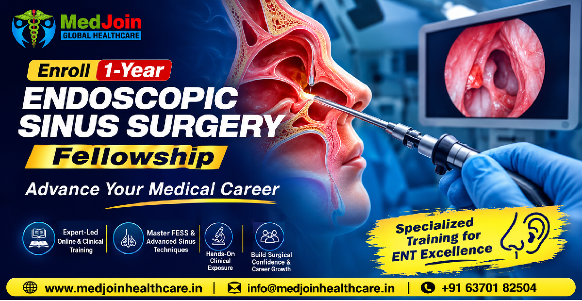
A 1-Year Fellowship in Endoscopic Sinus Surgery is designed for medical professionals who want to strengthen their expertise in advanced rhinology and develop greater confidence in the evaluation and management of patients requiring endoscopic sinus procedures.
The fellowship provides an opportunity to move beyond theoretical learning and understand how clinical decision-making, diagnostic interpretation, surgical planning, operative techniques, postoperative care, and patient safety come together in real-world ENT practice.
For doctors looking to advance their medical career, an organized fellowship can provide structured exposure to advanced concepts and help bridge the gap between postgraduate knowledge and focused subspecialty practice.
Whether you are an ENT specialist seeking focused training in rhinology or a medical professional looking to expand your understanding of endoscopic sinonasal procedures, a structured one-year fellowship can offer an academically oriented pathway for professional development.
What Is Endoscopic Sinus Surgery?
Endoscopic Sinus Surgery is a minimally invasive surgical approach used to diagnose and treat selected disorders affecting the nasal cavity and paranasal sinuses. Instead of relying primarily on large external surgical approaches, endoscopic techniques use specialized instruments and a camera system to provide visualization through the natural nasal passages.
Modern endoscopic sinus surgery is based on detailed knowledge of sinonasal anatomy and the functional relationship between the nasal cavity, sinus ostia, mucosa, and surrounding structures.
The primary objective is generally to restore or improve sinus ventilation and drainage while preserving healthy mucosa whenever clinically appropriate.
Endoscopic techniques have become an important part of contemporary rhinology because they can provide excellent visualization while allowing surgeons to address pathology through minimally invasive access.
However, successful endoscopic surgery requires much more than familiarity with an endoscope. A surgeon must understand three-dimensional anatomy, recognize anatomical variations, interpret imaging findings, select appropriate patients, anticipate surgical risks, use instruments safely, and manage postoperative healing.
This is why focused education in Endoscopic Sinus Surgery can be valuable for ENT professionals who want to develop advanced expertise.
Why Choose a Fellowship in Endoscopic Sinus Surgery?
ENT practice encompasses a wide range of conditions involving the ear, nose, throat, head, and neck. Rhinology itself is a highly specialized field, and advances in imaging, endoscopic equipment, navigation systems, instrumentation, and surgical techniques continue to influence clinical practice.
A dedicated fellowship allows learners to concentrate on one important area rather than studying rhinology only as a small component of a broader curriculum.
Key reasons to consider a fellowship include:
- Focused learning in rhinology and sinonasal disease
- Advanced understanding of nasal and paranasal sinus anatomy
- Structured study of endoscopic techniques
- Improved interpretation of CT imaging
- Better understanding of surgical indications
- Exposure to operative planning principles
- Understanding of endoscopic instruments and equipment
- Learning principles of perioperative management
- Recognition and prevention of complications
- Development of evidence-based clinical decision-making
- Opportunity to strengthen professional credentials
- Exposure to contemporary developments in rhinology
A fellowship should not simply be viewed as another certificate. Its value depends on the quality of its curriculum, faculty, clinical exposure, academic structure, assessment process, and practical learning opportunities.
For that reason, prospective candidates should carefully evaluate the institution, faculty, clinical environment, curriculum, accreditation or affiliation claims, assessment methods, and practical training arrangements before enrollment.
Who Can Benefit From a 1-Year Endoscopic Sinus Surgery Fellowship?
A focused fellowship may be particularly useful for doctors who already have foundational medical or ENT knowledge and want to develop a deeper understanding of endoscopic sinonasal surgery.
Potential candidates may include:
ENT Specialists
ENT specialists can use focused fellowship education to deepen their knowledge of rhinology and endoscopic sinus procedures.
ENT Postgraduates
Doctors who have completed relevant postgraduate training may seek additional structured exposure to advanced rhinology concepts.
Early-Career ENT Practitioners
Doctors beginning independent practice may benefit from systematic revision of anatomy, diagnostics, surgical planning, and postoperative management.
Doctors Expanding Their Clinical Scope
Professionals who want to develop a focused interest in rhinology can use a fellowship as an organized learning pathway.
Academically Interested Doctors
Those interested in teaching, clinical research, presentations, publications, and evidence-based medicine may benefit from a curriculum that integrates academic learning with clinical concepts.
Eligibility requirements vary according to the institution and program. Candidates should verify the exact eligibility criteria before applying.
Understanding the Importance of Sinonasal Anatomy
A strong understanding of anatomy is fundamental to safe Endoscopic Sinus Surgery.
The nasal cavity and paranasal sinuses form a complex anatomical region containing several important structures located close to the orbit, skull base, optic nerve, internal carotid artery, lacrimal drainage system, and other critical anatomical landmarks.
The paranasal sinuses generally include:
- Maxillary sinuses
- Frontal sinuses
- Ethmoid air cells
- Sphenoid sinuses
Each sinus has unique anatomical relationships, drainage pathways, and surgical considerations.
An endoscopic surgeon needs to understand not only the appearance of these structures but also their three-dimensional relationships.
Important anatomical concepts include:
- Nasal septum
- Inferior turbinate
- Middle turbinate
- Superior turbinate
- Uncinate process
- Ethmoid bulla
- Hiatus semilunaris
- Ethmoidal infundibulum
- Middle meatus
- Frontal recess
- Sphenoethmoidal recess
- Natural sinus ostia
- Skull base
- Lamina papyracea
- Orbit
- Optic nerve
- Internal carotid artery
A structured fellowship can provide repeated exposure to these concepts through lectures, imaging discussions, endoscopic demonstrations, case reviews, and practical learning where available.
The Role of Diagnostic Evaluation
Endoscopic sinus surgery should be based on appropriate clinical evaluation rather than imaging alone.
A comprehensive assessment may include:
- Detailed medical history
- Symptom evaluation
- Nasal examination
- Endoscopic assessment
- Relevant laboratory investigations
- Imaging when clinically indicated
- Evaluation of associated conditions
- Assessment of previous treatment
- Identification of surgical indications
- Discussion of expected outcomes and risks
The ability to integrate clinical findings with objective evidence is an essential component of advanced rhinology practice.
A fellowship can help doctors develop a structured approach to clinical reasoning so that surgical intervention is considered when appropriate rather than simply because an abnormality appears on imaging.
Nasal Endoscopy in Modern Rhinology
Nasal endoscopy is central to contemporary rhinology.
Endoscopic examination can provide direct visualization of structures that may not be adequately assessed through routine anterior rhinoscopy.
Depending on the clinical situation, endoscopy may help identify:
- Mucosal edema
- Polyps
- Purulent secretions
- Anatomical variations
- Septal abnormalities
- Masses
- Crusting
- Postoperative changes
- Signs of inflammation
- Obstruction around sinus drainage pathways
Learning systematic endoscopic examination is therefore an important component of focused rhinology education.
Doctors may study the principles of endoscope handling, orientation, visualization, documentation, interpretation, and clinical correlation.
Understanding Chronic Rhinosinusitis
Chronic rhinosinusitis is one of the major conditions encountered in rhinology practice.
It is characterized by persistent inflammation involving the nose and paranasal sinuses and may present with symptoms such as nasal obstruction, nasal discharge, facial pressure or pain, and reduced sense of smell.
Management depends on the patient's clinical presentation, disease phenotype, associated conditions, previous treatments, and objective findings.
Medical treatment may be appropriate in many patients, while surgery may be considered for selected patients who continue to have clinically significant disease despite appropriate medical management.
A fellowship curriculum can explore:
- Pathophysiology of chronic rhinosinusitis
- Clinical phenotypes
- Diagnostic criteria
- Nasal endoscopy
- CT interpretation
- Medical management
- Indications for surgery
- Surgical planning
- Postoperative care
- Recurrence and long-term management
Nasal Polyposis and Endoscopic Management
Nasal polyps are another important component of rhinology practice.
Patients may experience:
- Nasal obstruction
- Reduced smell
- Nasal discharge
- Facial pressure
- Mouth breathing
- Sleep disturbance
- Reduced quality of life
Nasal polyposis can be associated with chronic inflammatory disease and may coexist with conditions such as asthma or aspirin-exacerbated respiratory disease.
Management may involve medical therapy, surgery, advanced therapies in selected settings, and long-term follow-up.
Endoscopic sinus surgery can play an important role in appropriately selected patients.
A fellowship can help learners understand patient selection, disease assessment, surgical objectives, recurrence considerations, and postoperative management.
CT Scan Interpretation in Endoscopic Sinus Surgery
High-quality imaging interpretation is an essential skill for modern endoscopic sinus surgery.
CT imaging provides important information regarding:
- Sinus opacification
- Anatomical variations
- Osteomeatal complex
- Frontal recess
- Ethmoid anatomy
- Sphenoid anatomy
- Skull-base relationships
- Orbital anatomy
- Previous surgical changes
- Extent of disease
The surgeon must be able to correlate CT findings with endoscopic and clinical findings.
A fellowship may therefore include systematic training in reading and discussing CT scans.
Rather than memorizing individual radiological patterns, learners should develop the ability to ask clinically relevant questions:
- Where is the disease located?
- What is the likely anatomical pathway involved?
- Are there important anatomical variations?
- What structures are immediately adjacent to the surgical field?
- Has the patient undergone previous surgery?
- Are there features that may increase surgical risk?
- Does imaging correlate with the patient's symptoms and endoscopic findings?
This approach promotes safer and more thoughtful surgical planning.
Functional Principles of Endoscopic Sinus Surgery
Modern endoscopic sinus surgery emphasizes restoration of sinus function while avoiding unnecessary tissue disruption.
The principles may include:
- Identifying the natural drainage pathways
- Removing clinically significant obstruction
- Preserving healthy mucosa where possible
- Maintaining appropriate anatomical orientation
- Addressing the pathology responsible for symptoms
- Avoiding unnecessary surgical trauma
- Protecting adjacent critical structures
The exact surgical approach varies according to disease, anatomy, previous surgery, surgeon expertise, and patient-specific factors.
A fellowship can help doctors understand these principles rather than simply memorizing procedural steps.
Endoscopic Maxillary Sinus Surgery
The maxillary sinus is one of the most commonly encountered paranasal sinuses in rhinology.
Endoscopic approaches may be used for selected inflammatory disease, chronic sinus disease, certain benign lesions, and other appropriately evaluated conditions.
Training may cover:
- Relevant maxillary sinus anatomy
- Natural ostium
- Uncinate process
- Ethmoid infundibulum
- Middle meatus
- Surgical indications
- Endoscopic visualization
- Instrumentation
- Postoperative care
- Potential complications
Understanding the relationship between the maxillary sinus and surrounding structures is important for safe intervention.
Ethmoid Sinus Surgery
The ethmoid region is anatomically complex and particularly important in endoscopic sinus surgery.
The ethmoid labyrinth contains multiple air cells with close relationships to the orbit and skull base.
A structured educational program may explore:
- Anterior ethmoid anatomy
- Posterior ethmoid anatomy
- Ethmoid bulla
- Lamina papyracea
- Skull base
- Middle turbinate
- Basal lamella
- Endoscopic orientation
- Surgical planning
- Complication avoidance
Because of the complexity of this region, detailed anatomical understanding is critical.
Frontal Sinus Surgery
The frontal sinus and frontal recess represent one of the more challenging areas of endoscopic sinus surgery.
The anatomy can vary substantially between patients, making individualized imaging review and careful surgical planning particularly important.
Fellowship education may cover:
- Frontal sinus anatomy
- Frontal recess
- Agger nasi region
- Frontal cells
- Imaging interpretation
- Surgical approaches
- Endoscopic orientation
- Postoperative surveillance
- Management of restenosis and recurrent disease
Advanced frontal sinus surgery requires a strong understanding of three-dimensional anatomy and careful risk assessment.
Sphenoid Sinus Surgery
The sphenoid sinus has important anatomical relationships with structures including the optic nerve, internal carotid artery, cavernous sinus, and skull base.
This makes knowledge of sphenoid anatomy particularly important.
Educational topics may include:
- Sphenoid sinus anatomy
- Sphenoid ostium
- Septations
- Optic nerve relationship
- Carotid artery relationship
- Skull-base anatomy
- Endoscopic orientation
- Surgical planning
- Complication prevention
The fellowship should emphasize careful anatomical identification and patient-specific planning.
Advanced Endoscopic Techniques
As expertise develops, doctors may encounter increasingly complex sinonasal cases.
Advanced topics may include:
- Revision sinus surgery
- Difficult frontal recess anatomy
- Extensive polyposis
- Fungal sinus disease
- Selected sinonasal masses
- CSF leak principles
- Orbital complications
- Skull-base considerations
- Image-guided surgery
- Multidisciplinary management
Advanced techniques should be taught within an appropriate clinical and institutional framework.
Image-Guided Surgery
Image-guided navigation has become an important adjunct in selected complex sinonasal procedures.
Navigation systems can help surgeons correlate the operative field with preoperative imaging.
Potential applications may include selected:
- Revision procedures
- Extensive inflammatory disease
- Anatomically complex cases
- Frontal sinus procedures
- Skull-base-related surgery
- Lesions near critical structures
However, navigation is an adjunct and does not replace detailed anatomical knowledge or surgical judgment.
A fellowship can introduce learners to the principles, indications, limitations, and workflow associated with image-guided surgery.
Endoscopic Management of Fungal Sinus Disease
Fungal disease of the sinonasal tract encompasses different clinical entities, ranging from non-invasive conditions to invasive disease.
The clinical presentation and treatment strategy can vary significantly.
A focused curriculum may cover:
- Fungal sinus disease classification
- Clinical presentation
- Imaging features
- Endoscopic findings
- Diagnostic considerations
- Medical management
- Surgical principles
- Histopathology
- Multidisciplinary care
- Postoperative monitoring
Invasive fungal disease can represent a medical emergency, requiring urgent specialist assessment and multidisciplinary management.
Sinonasal Tumors and Endoscopic Approaches
Endoscopic techniques have expanded the surgical options for selected sinonasal lesions and tumors.
However, tumor management requires careful diagnosis, staging, imaging, pathology, multidisciplinary planning, and appropriate surgical expertise.
Fellowship education may introduce:
- Common sinonasal tumors
- Clinical presentation
- Endoscopic examination
- Imaging
- Biopsy principles
- Histopathological correlation
- Surgical planning
- Multidisciplinary management
- Follow-up
Candidates should understand that complex oncological cases require specialized institutional expertise and should not be managed solely on the basis of fellowship-level theoretical training.
CSF Leak: An Important Rhinology Topic
Cerebrospinal fluid leaks can occur spontaneously, after trauma, or as a complication of surgery.
Recognition and appropriate management are critical because persistent CSF leakage can increase the risk of serious complications.
A fellowship may cover:
- Recognition of suspected CSF leak
- Relevant skull-base anatomy
- Diagnostic principles
- Imaging
- Endoscopic repair concepts
- Graft and reconstruction principles
- Postoperative monitoring
- Complication management
This is an area where practical exposure under experienced specialists can be particularly valuable.
Orbital Complications
The orbit is closely related to the paranasal sinuses, particularly the ethmoid region.
Potential complications can include:
- Orbital injury
- Hemorrhage
- Diplopia
- Extraocular muscle injury
- Visual impairment
- Orbital hematoma
A comprehensive fellowship should emphasize anatomical knowledge, recognition of risk, prevention, early identification of complications, and appropriate escalation of care.
Patient safety should remain the primary objective of every surgical training program.
Skull-Base Safety
The skull base is another critical anatomical region in endoscopic sinus surgery.
Potential complications involving the skull base can be serious.
Therefore, training should emphasize:
- Identification of skull-base anatomy
- CT-based surgical planning
- Anatomical variations
- Recognition of high-risk areas
- Controlled instrumentation
- Prevention of excessive tissue trauma
- Recognition of CSF leak
- Appropriate management and referral
A structured fellowship can help reinforce the importance of surgical discipline and anatomical orientation.
Perioperative Patient Management
Endoscopic sinus surgery does not begin when the patient enters the operating room.
Good outcomes depend on appropriate preoperative assessment, informed consent, optimization of relevant medical conditions, surgical planning, postoperative treatment, and follow-up.
A fellowship may address:
Preoperative Care
- Medical history
- Medication review
- Relevant comorbidities
- Allergy history
- Previous surgery
- Endoscopic assessment
- Imaging
- Informed consent
Intraoperative Considerations
- Patient positioning
- Endoscopic visualization
- Instrumentation
- Hemostasis
- Tissue preservation
- Critical structure identification
- Communication with the surgical team
Postoperative Care
- Nasal care
- Endoscopic follow-up
- Debridement where clinically indicated
- Medical therapy
- Infection monitoring
- Bleeding assessment
- Recurrence surveillance
Understanding Surgical Instruments
Familiarity with endoscopic instruments is an important part of practical learning.
Depending on the clinical environment, trainees may encounter:
- Rigid nasal endoscopes
- Angled endoscopes
- Suction instruments
- Blakesley forceps
- Through-cutting instruments
- Microdebriders
- Balloon systems
- Navigation equipment
- Hemostatic devices
- Powered instruments
Understanding the purpose and limitations of equipment helps promote efficient and safe surgical practice.
Hands-on equipment training should always occur under appropriate supervision.
The Role of Simulation-Based Training
Simulation can complement clinical learning.
Possible simulation methods include:
- Endoscopic anatomy models
- Cadaveric dissection where available
- Virtual surgical platforms
- Instrument handling exercises
- CT-to-anatomy correlation
- Surgical planning exercises
- Case-based simulations
Simulation allows learners to develop familiarity with anatomy and procedural concepts before performing clinical tasks.
It is particularly useful for practicing orientation, instrument handling, surgical planning, and complication recognition in a controlled environment.
Hands-On Clinical Training
One of the most valuable components of a clinical fellowship can be supervised practical exposure.
A hands-on component may provide opportunities to observe or participate in appropriate activities according to the learner's qualifications, institutional policies, and supervising faculty.
Clinical exposure may include:
- Outpatient ENT clinics
- Nasal endoscopy
- Preoperative evaluation
- CT review
- Surgical observation
- Operating-room exposure
- Postoperative follow-up
- Case discussions
- Multidisciplinary meetings
The exact level of procedural participation should always depend on credentialing, competence, supervision, institutional policy, and applicable regulations.
Online Learning and Clinical Fellowship
A modern fellowship can combine digital education with clinical learning.
An online component may provide:
- Recorded lectures
- Reading materials
- Case discussions
- Video demonstrations
- Assignments
- MCQ assessments
- Live academic sessions
- Clinical case reviews
Digital learning offers flexibility for working doctors.
However, online education alone cannot substitute for supervised clinical experience when the intended learning outcomes involve practical procedural skills.
A strong hybrid structure should clearly distinguish theoretical learning from hands-on competency development.
A Typical 12-Month Learning Journey
A one-year fellowship can be organized into progressive stages.
Months 1–3: Foundations
The initial phase may focus on:
- Sinonasal anatomy
- Physiology
- Pathophysiology
- Diagnostic evaluation
- Nasal endoscopy
- CT interpretation
- Common rhinological disorders
Months 4–6: Core Endoscopic Surgery
The middle phase may introduce:
- Surgical indications
- Endoscopic principles
- Maxillary sinus surgery
- Ethmoid surgery
- Frontal sinus approaches
- Sphenoid sinus approaches
- Instrumentation
- Perioperative care
Months 7–9: Advanced Rhinology
Learners may explore:
- Revision surgery
- Difficult anatomy
- Nasal polyposis
- Fungal disease
- Image-guided surgery
- Selected sinonasal lesions
- Complication management
Months 10–12: Integration and Advanced Case Learning
The final phase may emphasize:
- Comprehensive case planning
- Complex case discussions
- Surgical decision-making
- Postoperative management
- Academic presentations
- Research or audit projects
- Final assessment
The actual curriculum and clinical exposure will depend on the fellowship provider.
Evidence-Based Medicine in Rhinology
Modern clinical practice should be based on the best available evidence combined with clinical expertise and patient preferences.
A fellowship can introduce learners to:
- Clinical guidelines
- Systematic reviews
- Meta-analysis
- Randomized trials
- Observational studies
- Critical appraisal
- Research methodology
- Statistical interpretation
Doctors should learn to distinguish high-quality evidence from anecdotal claims.
This is particularly important in surgical specialties where techniques can evolve rapidly.
Career Benefits of an Endoscopic Sinus Surgery Fellowship
A focused fellowship may contribute to professional development in several ways.
1. Specialized Knowledge
The program can deepen knowledge in rhinology and endoscopic surgery.
2. Clinical Confidence
Structured exposure to cases may help doctors approach relevant clinical situations more systematically.
3. Academic Development
Assignments, presentations, case discussions, and research can strengthen academic skills.
4. Professional Profile
A recognized and appropriately documented fellowship may add a focused qualification to a doctor's professional portfolio, subject to the program's accreditation and recognition.
5. Networking
Interaction with faculty and other clinicians can support professional networking.
6. Continuing Professional Development
The fellowship can provide structured learning for doctors committed to lifelong education.
Hybrid vs Hands-On Fellowship
Prospective candidates often compare online, hybrid, and hands-on programs.
Hybrid Fellowship
A hybrid model can combine online education with clinical exposure.
Potential advantages include:
- Flexibility
- Access to digital resources
- Reduced travel requirements
- Structured theoretical learning
- Selected clinical exposure
Hands-On Fellowship
A predominantly clinical fellowship may provide greater direct exposure to hospital-based learning.
Potential advantages include:
- Regular clinical interaction
- Operating-room exposure
- Direct observation
- Supervised procedural learning
- Postoperative follow-up
The best format depends on the learner's goals, location, clinical availability, and professional commitments.
Candidates should carefully confirm exactly what "hands-on" means in a particular program.
Artificial Intelligence and Rhinology
Artificial intelligence is increasingly being explored in medical imaging and clinical decision support.
Potential applications in rhinology may include:
- Image analysis
- Disease classification
- Automated measurements
- Surgical planning support
- Research
- Patient outcome prediction
However, AI-generated recommendations should be interpreted cautiously and validated clinically.
Doctors should understand both the potential and limitations of emerging technologies.
A Career-Oriented Learning Experience
For a doctor, professional development is not only about acquiring additional knowledge. It is about becoming more systematic, evidence-based, confident, and patient-centered.
A well-designed one-year fellowship can create an organized framework for this development.
It can bring together:
Anatomy + Diagnostics + Imaging + Clinical Reasoning + Surgical Principles + Practical Exposure + Research + Assessment
This integrated approach is particularly valuable in a technically demanding field such as rhinology.
Why Choose MedJoin Global Healthcare for Fellowship Education?
MedJoin Global Healthcare offers structured medical fellowship programs designed to support continuing professional education for healthcare professionals.
For candidates considering an Endoscopic Sinus Surgery Fellowship, it is important to review the specific program curriculum, faculty, training arrangements, eligibility criteria, assessment process, certification details, and applicable accreditation or affiliations before enrollment.
The objective should be to choose a program that aligns with your professional goals and provides meaningful academic and clinical learning.
Visit www.medjoinhealthcare.in to explore the available fellowship programs and obtain current program information.
Prospective candidates should independently verify all accreditation, affiliation, certification, clinical-training, and career-related claims before making an enrollment decision.
Your Next Step Toward Advanced Rhinology Learning
Medical careers evolve through education, experience, mentorship, and continuous improvement.
A 1-Year Endoscopic Sinus Surgery Fellowship can provide a structured pathway for doctors who want to develop focused knowledge in rhinology and endoscopic sinus surgery.
From anatomy and diagnostics to surgical principles, imaging, clinical case discussions, complication management, postoperative care, and academic development, the fellowship journey can help learners build a stronger foundation for continued professional growth.
Endoscopic Sinus Surgery represents one of the most important developments in modern rhinology. Its minimally invasive approach, advanced visualization, and ability to address selected sinonasal disorders have made endoscopic techniques an integral part of contemporary ENT practice.
Yet successful endoscopic surgery requires much more than technical familiarity.
It requires a detailed understanding of anatomy, careful diagnosis, appropriate patient selection, CT interpretation, surgical planning, instrumentation, complication prevention, postoperative management, and lifelong learning.
A 1-Year Endoscopic Sinus Surgery Fellowship can provide a structured educational environment for doctors seeking focused professional development in this field.
For aspiring rhinology-focused clinicians, the journey should begin with strong fundamentals and progress toward advanced clinical reasoning, supervised practical exposure, evidence-based decision-making, and responsible patient care.
Advance your medical career. Strengthen your rhinology knowledge. Build your expertise in Endoscopic Sinus Surgery.
Explore the 1-Year Endoscopic Sinus Surgery Fellowship and take the next step in your professional learning journey.